LOW INTENSITY PULSED ULTRASOUND ATTENUATES GLOMERULAR ENDOTHELIAL DYSFUNCTION BY AMELIORATING MITOCHONDRIAL DAMAGE AND CELLULAR SENESCENCE IN DIABETIC NEPHROPATHY


Certificate Output Instructions

For best output, select "Paper Size" as "A4" and "Margin" as "0" or "None".

To save or print to PDF, please select Print Destination > Save as PDF, enable Background Graphics under "More Settings", then click "Save".


Certificate Background


Presented the abstract " "
(Abstract co-author(s):  )


E-Poster Presentation

During the congress, E-Posters will be accessible to all participants on the congress website 24/7, as well as in the E-poster stations in the congress center. 

Preparing your E-Poster

Please review the E-Poster format requirements carefully when preparing your E-Poster. Should your E-Poster not meet the mentioned requirements, it may not be displayed as described above.

​E-Poster Submission Deadline

Please prepare and upload your E-Poster no later than March 14, 2026 11.59PM CET. After this date, you will no longer be able to prepare and upload your E-poster and it will not be displayed and accessible on the congress website.​

E-Poster Format Requirements
  • PDF file
  • Layout: Portrait (vertical orientation)
  • One page only (Dim A4: 210 x 297mm or PPT)
  • E-Poster can be prepared in PowerPoint (one (1) PowerPoint slide) but must be saved and submitted as PDF file.
  • File Size: Maximum file size is 2 Megabytes (2 MB)
  • No hyperlinks, animated images, animations, and slide transitions
  • Language: English
  • Include your abstract number
  • E-posters can include QR codes, tables and photos
https://storage.unitedwebnetwork.com/files/1099/a7ee8bbd09852fb1cb6ac71477a9800a.pdf
LOW INTENSITY PULSED ULTRASOUND ATTENUATES GLOMERULAR ENDOTHELIAL DYSFUNCTION BY AMELIORATING MITOCHONDRIAL DAMAGE AND CELLULAR SENESCENCE IN DIABETIC NEPHROPATHY

Please follow the instructions below to input your abstract title.

Abstract titles should be brief and reflect the content of the abstract.

  • The title will not be accepted if it exceeds 25 words.
  • Type in CAPITAL LETTERS.
  • Lowercase may be used for abbreviations only, for example, mRNA.
Yuankang
Zhu
Yuankang Zhu 15258683336@sjtu.edu.cn Xinhua Hospital Affiliated to Shanghai Jiao Tong University School of Medicine Department of Gerontology Shanghai China *
Liangshi Hao haoliangshi@outlook.com Shanghai Ninth People's Hospital Affiliated to Shanghai Jiaotong University School of Medicine Department of Urology Shanghai China -
Shengzhuo You youshengzhuo2001@163.com Zhongshan Hospital Affiliated to Fudan University Department of Nephrology Shanghai China -
Changning Hao gilberthaocn@163.com Xinhua Hospital Affiliated to Shanghai Jiao Tong University School of Medicine Department of Gerontology Shanghai China -
Junli Duan duanjunli@xinhuamed.com.cn Xinhua Hospital Affiliated to Shanghai Jiao Tong University School of Medicine Department of Gerontology Shanghai China -
Yiqin Shi shiyiqin@zs-hospital.sh.cn Zhongshan Hospital Affiliated to Fudan University Department of Nephrology Shanghai China -


Diabetic Nerphropathy (DN) is a leading cause of end-stage renal disease. However, despite strict glycemic control through lifestyle interventions and pharmacotherapy, halting or reversing its progression remains challenging, highlighting the urgent needs for novel, non-glycemia-dependent therapeutic strategies. The glomerular endothelial cell (GEC), which serves as the first barrier sensing metabolic disturbances and hemodynamic abnormalities, plays a pivotal role in the initiation and progression of DN. Nonetheless, there is a notable lack of effective clinical therapies that protect GEC function. Therefore, developing novel therapies capable of directly improving GEC damage is of paramount importance. Low-Intensity Pulsed Ultrasound (LIPUS), a non-invasive physical therapy with broad applications in various disease models. Therefore, our present work aims to investigate whether LIPUS can ameliorate glomerular endothelial dysfunction in DN.

Type 2 diabetes mellitus (T2DM) models were established in C57BL/6J mice through 8 weeks of high-fat diet (HFD) feeding followed by intraperitoneal injections of streptozotocin (STZ, 40 mg/kg). DN mice were then randomly assigned to receive either LIPUS treatment (0.2 W/cm², 20% duty cycle, 1 MHz, 9 minutes every other day) or sham treatment for 12 weeks (Figure 1A). Renal function was assessed using urine samples collected from 12-hour metabolic cage studies and serum biochemical parameters. The efficacy of LIPUS treatment on GEC injury was evaluated through multi-omics analyses, histopathological examination, transmission electron microscopy, and molecular experiments.

Figure 1 LIPUS treatment improves renal function in DN mice. (A) Schematic of the LIPUS treatment protocol in DN mice. (B) Body weight were recorded weekly (n=6). (C) Blood glucose levels were monitored every two weeks. (n=6). (D) Renal index ratio of mice (n=6). (E) Blood urea nitrogen and serum creatinine (n=6). (F) Twelve hours urinary creatinine clearance, urinary albumin and urinary albumin-creatinine ratio (n=6). (G) The levels of CHO, HDL, LDL, and TG in mice serum (n=4). (H) Insulin tolerance was measured by ITT (n=4). (I) The area under curve of 120 minutes in ITT test. Data are presented as mean ± SD.  *P < 0.05, **P < 0.01, ***P < 0.001, and #P < 0.0001. LIPUS, Low-Intensity Pulsed Ultrasound; DN, Diabetic Nephropathy; ITT, Insulin Tolerance Test; BUN, Blood Urea Nitrogen; Scr, Serum Creatinine; UACR, Urinary Albumin-to-Creatinine Ratio; CHO, total cholesterol; HDL, high-density lipoprotein cholesterol; LDL, low-density lipoprotein cholesterol; TG, triglycerides; AUC, area under curve.Figure 2 LIPUS treatment attenuates GEC damage in DN mice. (A) Representative H&E, Masson, PAS-stained and CD31 (green) immunofluorescence images of mouse kidneys. Scale bar, 50 um. (B) Quantification of average collagen positive area (n=4). (C) Quantification of mesangial matrix fraction (n=4). (D) Quantification of glomerular area (n=4). (E) Quantitative analysis of the CD31 fluorescence intensity (n=4). (F) Representative western blot images of p-enos, t-enos, VCAM-1 and ET-1 in renal cortex. (G) Quantitative analysis of p- eNOS protein expression normalized to t-eNOS (n=6). (H) Quantitative analysis of Vcam-1 protein expression normalized to GAPDH (n=6). (I) Quantitative analysis ET-1 protein expression normalized to GAPDH (n=6).  Data are presented as mean ± SD.  *P < 0.05, **P < 0.01, ***P < 0.001, and #P < 0.0001. LIPUS, Low-Intensity Pulsed Ultrasound; GEC, glomerular endothelial cell; DN, Diabetic Nephropathy; H&E, Hematoxylin and Eosin; PAS, Periodic Acid–Schiff.Figure 3. LIPUS treatment ameliorates mitochondrial damage in GEC. (A) Schematic workflow of the multi-omics analysis performed on the renal cortex. (B) Venn diagram identifying the common DEGs from the RNA-seq and proteomic analyses. (C) GO functional enrichment analysis of the common DEGs. (D) KEGG pathway enrichment analysis of the common DEGs. (E) Representative TEM images of GEC in the mouse renal cortex (n=4). Scale bar, 2 μm. (F) Quantitative analysis of GEC ultrastructure, mitochondrial density, and mitochondrial volume based on TEM images. (G) Representative western blot images of Bcl-2, Bax, total Caspase 3 and cleaved Caspase 3 in renal cortex. (H) Quantitative analysis of Bcl-2 protein expression normalized to Bax (n=6). (H) Quantitative analysis of cleaved Caspase 3 protein expression normalized to total Caspase 3 (n=6). Data are presented as mean ± SD.  *P < 0.05, **P < 0.01, ***P < 0.001, and #P < 0.0001. LIPUS, Low-Intensity Pulsed Ultrasound; GEC, glomerular endothelial cell; DN, Diabetic Nephropathy; DEG, Differentially Expressed Genes; GO, Gene Ontology; KEGG, Kyoto Encyclopedia of Genes and Genomes; TEM, Transmission Electron Microscopy.
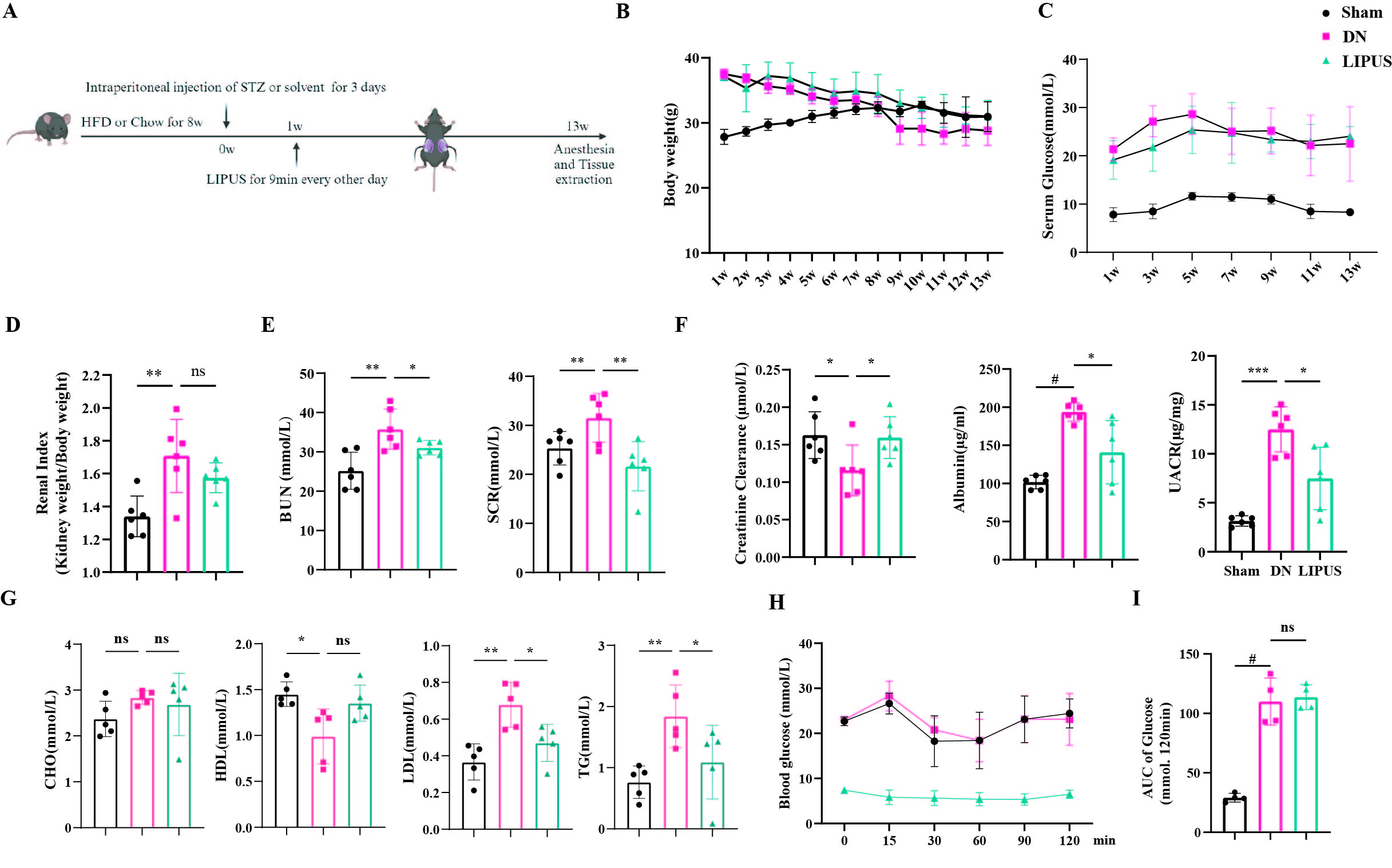
As diabetes progressed, mice in both the DN and LIPUS groups exhibited a continuous decrease in body weight, while blood glucose levels remained elevated (Figure 1B and 1C). As shown in Figure 1D, DN mice displayed renal hypertrophy, with a significantly higher renal index compared to the sham group, and LIPUS treatment only partially ameliorate this condition. The increased levels of blood urea nitrogen (BUN) and serum creatinine (SCR) in DN mice were downregulated after LIPUS intervention (Figure 1E). Furthermore, LIPUS treatment improved the lower creatinine clearance rate and attenuated the increases in urinary albumin levels and the urinary albumin-to-creatinine ratio in DN mice (Figure 1F). Notably, LIPUS also ameliorated lipid metabolism disorders associated in DN mice, including low-density lipoprotein (LDL) and triglyceride (TG) (Figure 1G). However, LIPUS treatment did not improve the characteristic insulin resistance features of T2DM (Figures 1H and 1I). These results suggest that the improvement of renal function by LIPUS in DN may be independent of blood glucose management.

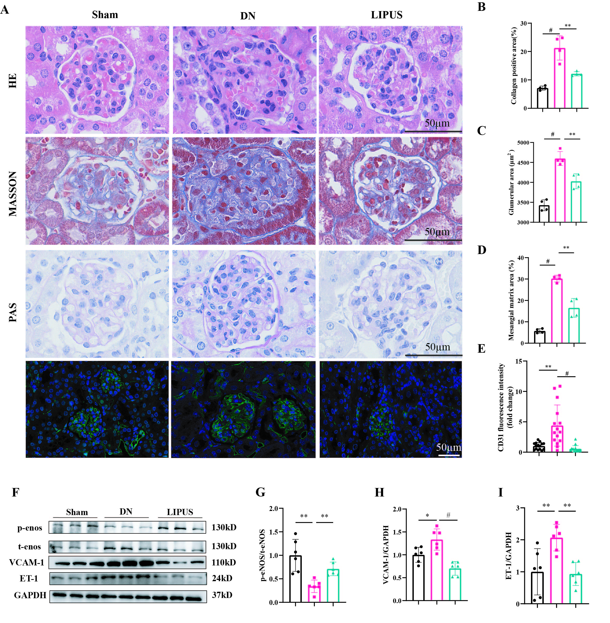
 Furthermore, histological examination was performed to evaluate the efficacy of LIPUS treatment. H&E staining and Masson’s trichrome staining indicated that LIPUS effectively reduced the extent of renal fibrosis compared with the DN group (Figures 2A and 2B). Periodic Acid–Schiff staining revealed that DN mice exhibited significant glomerular hypertrophy and mesangial matrix expansion, which were attenuated by LIPUS treatment (Figures 2A-D). CD31 staining was used to assess GEC injury. In the DN group, hypertrophic glomeruli were accompanied by abnormal vascular hyperplasia, which was ameliorated following LIPUS intervention (Figures 2A and 2E). We further examined markers associated with endothelial cell injury by Western blot. Compared with the sham group, the DN group showed a significant decrease in p-eNOS protein levels, along with increased expression of VCAM-1 and ET-1. In contrast, LIPUS treatment upregulated p-eNOS expression and downregulated the levels of VCAM-1 and ET-1 relative to the DN group (Figures 2F–2I). These findings suggest that LIPUS treatment may confer a protective effect against glomerular endothelial injury in DN.

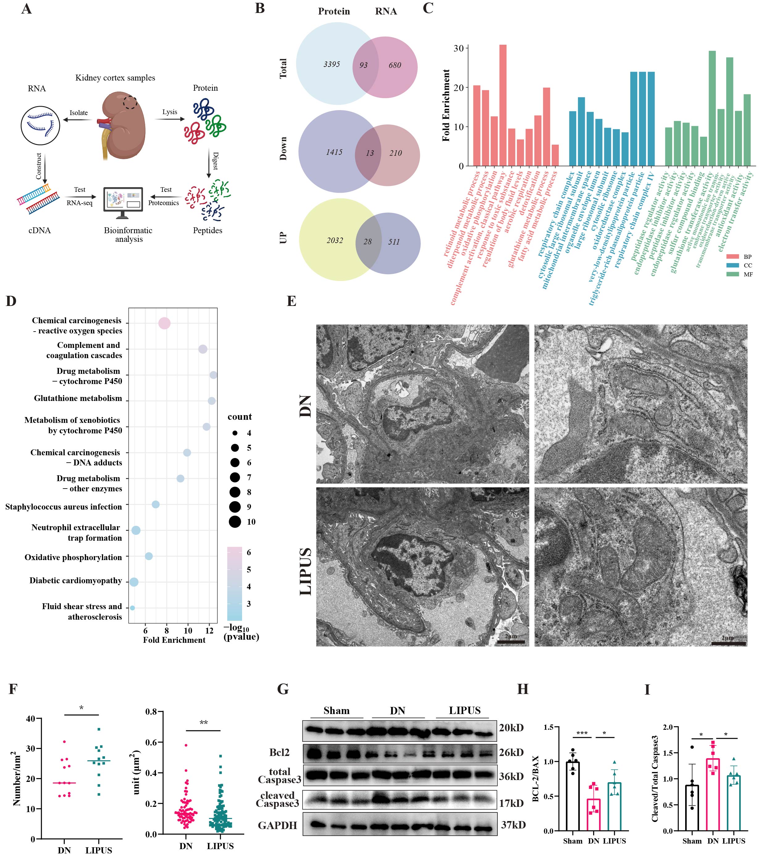
To further explore the underlying mechanisms and key targets of LIPUS treatment, we performed 4D label-free proteomic analysis and RNA sequencing on renal cortex samples from DN and LIPUS treated mice (Figure 3A). Venn diagram analysis identified 93 common differentially expressed genes (DEG) at the RNA and protein levels (Figure 3B). Among these overlapping genes, the number upregulated by LIPUS was significantly greater than those downregulated. Gene Ontology (GO) enrichment analysis revealed multiple terms significantly associated with mitochondrial processes, including oxidative phosphorylation, respiratory chain complex, mitochondrial intermembrane space, and oxidoreductase complex (Figure 3C). Similarly, KEGG pathway analysis indicated significant enrichment of mitochondria-related pathways such as oxidative stress, cytochrome P450 metabolism, and oxidative phosphorylation (Figure 3D). We next performed transmission electron microscopy to evaluate mitochondrial ultrastructure in GECs. TEM images showed that LIPUS treatment ameliorated the podocyte loss and glomerular structural damage induced by DN (Figure 3E). Mitochondria in the GECs of DN mice exhibited a significant reduction in number, accompanied by membrane disruption, structural disorganization, and shortened, fragmented, and blurred cristae. In contrast, LIPUS treatment effectively restored mitochondrial ultrastructure, increasing mitochondrial number and reducing mitochondrial volume (Figure 3F). Consistent with these morphological improvements, LIPUS upregulated the anti-apoptotic protein BCL2 and downregulated the pro-apoptotic proteins BAX and cleaved caspase-3, which are associated with the functions and integrity of mitochondrial. These findings suggest that LIPUS may alleviate GEC dysfunction and injury by attenuating mitochondrial damage.
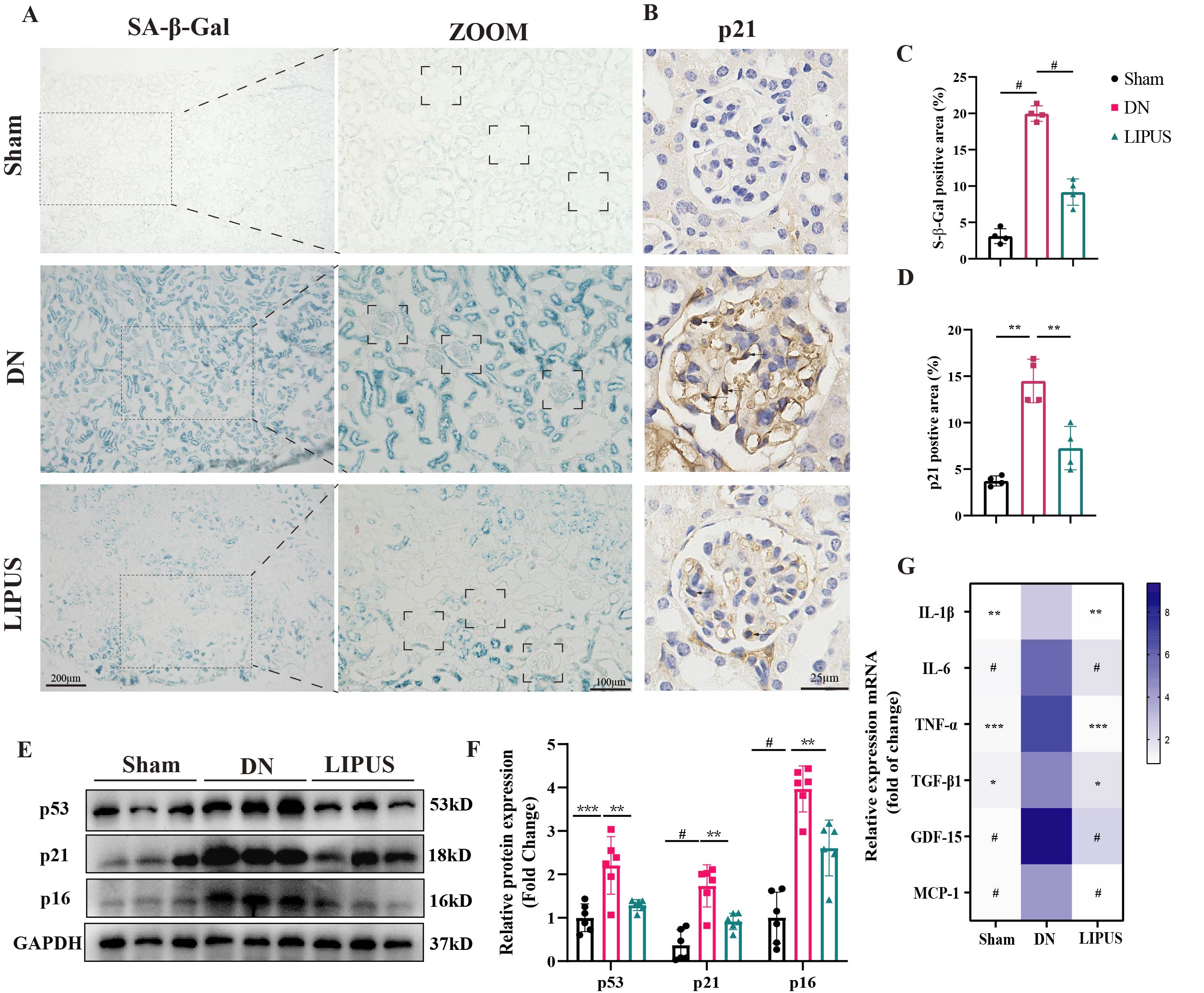
Given that mitochondrial dysfunction is a pivotal factor of cellular senescence, and GEC senescence has been established as a key pathological mechanism in diabetic nephropathy, we next assessed whether the amelioration of mitochondrial injury by LIPUS could subsequently attenuate the senescent phenotype in diabetic kidneys. As shown in Figure 4A, renal sections from the DN group exhibited a significantly larger SA-β-gal-positive area than those from the sham group, an effect markedly attenuated by LIPUS treatment, particularly within glomeruli (Figure 4C). Immunohistochemical staining for p21 in mouse glomeruli further indicated that LIPUS reduced its elevated expression in DN kidneys (Figure 4B and 4D). Consistent with these findings, Western blot analysis revealed that the protein levels of senescence markers p53, p21, and p16 were upregulated in DN kidneys compared with sham controls, and LIPUS treatment effectively suppressed this increase (Figure 4E and 4F). Similarly, the mRNA levels of key senescence-associated secretory phenotype (SASP) factors were significantly elevated in DN kidneys relative to the sham group, and LIPUS treatment also downregulated these SASP expression (Figure 4G).

This study demonstrates that LIPUS significantly improved renal dysfunction and GEC damage in DN without managing blood glucose. These beneficial effects are likely attributable to the restoration of mitochondrial structure and function, along with the downregulation of senescence-associated markers. Our findings reveal LIPUS as a novel glycemic control-independent therapeutic strategy, providing a promising direction for the clinical management of DN.

Kewords