CASE REPORT: A VERY YOUNG GIRL , 15-YEAR-OLD, WITH ISOLATED ANTI-GBM GLOMERULONEPHRITIS


Certificate Output Instructions

For best output, select "Paper Size" as "A4" and "Margin" as "0" or "None".

To save or print to PDF, please select Print Destination > Save as PDF, enable Background Graphics under "More Settings", then click "Save".


Certificate Background


Presented the abstract " "
(Abstract co-author(s):  )


E-Poster Presentation

During the congress, E-Posters will be accessible to all participants on the congress website 24/7, as well as in the E-poster stations in the congress center. 

Preparing your E-Poster

Please review the E-Poster format requirements carefully when preparing your E-Poster. Should your E-Poster not meet the mentioned requirements, it may not be displayed as described above.

​E-Poster Submission Deadline

Please prepare and upload your E-Poster no later than March 14, 2026 11.59PM CET. After this date, you will no longer be able to prepare and upload your E-poster and it will not be displayed and accessible on the congress website.​

E-Poster Format Requirements
  • PDF file
  • Layout: Portrait (vertical orientation)
  • One page only (Dim A4: 210 x 297mm or PPT)
  • E-Poster can be prepared in PowerPoint (one (1) PowerPoint slide) but must be saved and submitted as PDF file.
  • File Size: Maximum file size is 2 Megabytes (2 MB)
  • No hyperlinks, animated images, animations, and slide transitions
  • Language: English
  • Include your abstract number
  • E-posters can include QR codes, tables and photos
 
CASE REPORT: A VERY YOUNG GIRL , 15-YEAR-OLD, WITH ISOLATED ANTI-GBM GLOMERULONEPHRITIS

Please follow the instructions below to input your abstract title.

Abstract titles should be brief and reflect the content of the abstract.

  • The title will not be accepted if it exceeds 25 words.
  • Type in CAPITAL LETTERS.
  • Lowercase may be used for abbreviations only, for example, mRNA.
AUNG
ZAW HTET
AUNG ZAW HTET aungzawhtetdr@gmail.com Defence Services General Hospital Nephrology Yangon Myanmar *
-
-
-
-
-
-
-
-
-
-
-
-
-
-

Anti-glomerular basement membrane disease is a very rare small vessel vasculitis and most patients presented with rapidly progressive glomerulonephritis. This disorder is extremely severe and can progress to End-stage renal disease (ESRD).Anti-GBM disease is rare and its occurrence was fewer than two cases per million population. This disease has bimodal age distribution , with peak incidence in the third decade and in the sixth to seventh decades. This 15 years old girl was currently on hemodialysis after getting hit by isolated anti-GBM glomerulonephritis. She was treated with urgent initiation of hemodialysis in view of rapidly progressive renal failure (RPRF).


A very young girl (15 years old)  presented with generalized oedema and anuria for 2 days and confusion. On examination , GCS 3/15, BP-160/100 mmHg, PR-96/min, Heart – S1+S2+0, lungs- VBS with bibasal crepts, Abd- abdominal wall oedema, bilateral leg oedema (+), extensor plantar response on the left side . Urgent CT revealed acute infarct at right parietal lobe. 

 Urgent investigations revealed HBsAg, HCV Ab and HIV Ab- all clear, Urea – 31.32 mmol/L, Creatinine-1226 umol/L , Na-132, K-5.7, HCO3-19 , serum albumin- 23 g/dl, Hb-9.2 , WBC- 12.16 , Platelet – 285. 

Urgent hemodialysis was started with right IJV temporary HD sheath.

After first time hemodialysis, she got fully concious with GCS 15/15) and her vital signs were stable and urine was coming out.

ANA – 1:80 (Speckled pattern, Trace), ENA profile – All Negative,

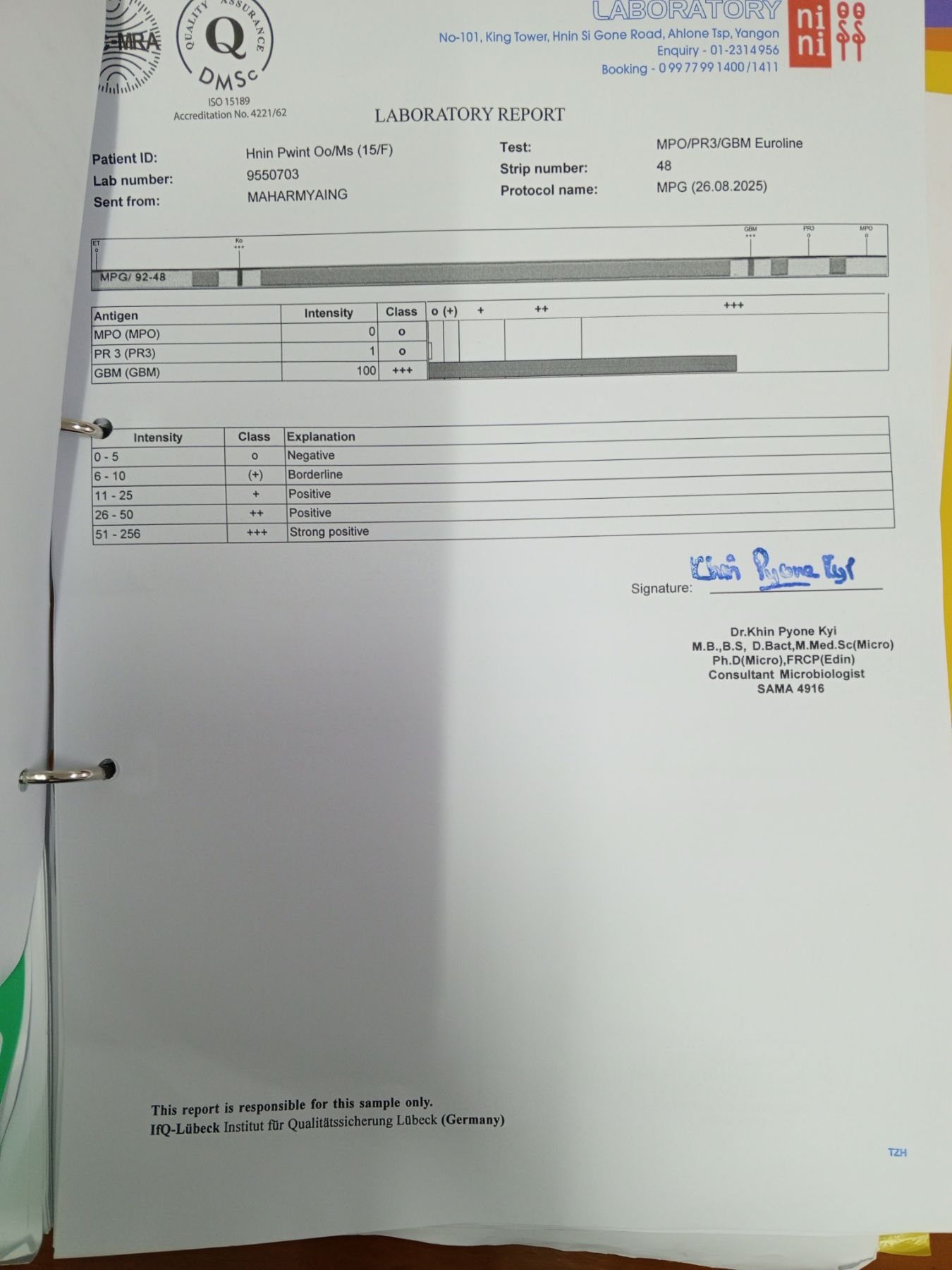
Anti PR3 – Negative, Anti MPO -Negative, Anti GBM (storongly Positive)- (+++) . 

USG (abd) revealed right kidney – 9.37 cm with cortical thickness – 1.7cm , Left Kidney -10.85 cm with cortical thickness 1.62 cm, Ascites (++) and bilateral pleural effusion and stomach wall oedema. 

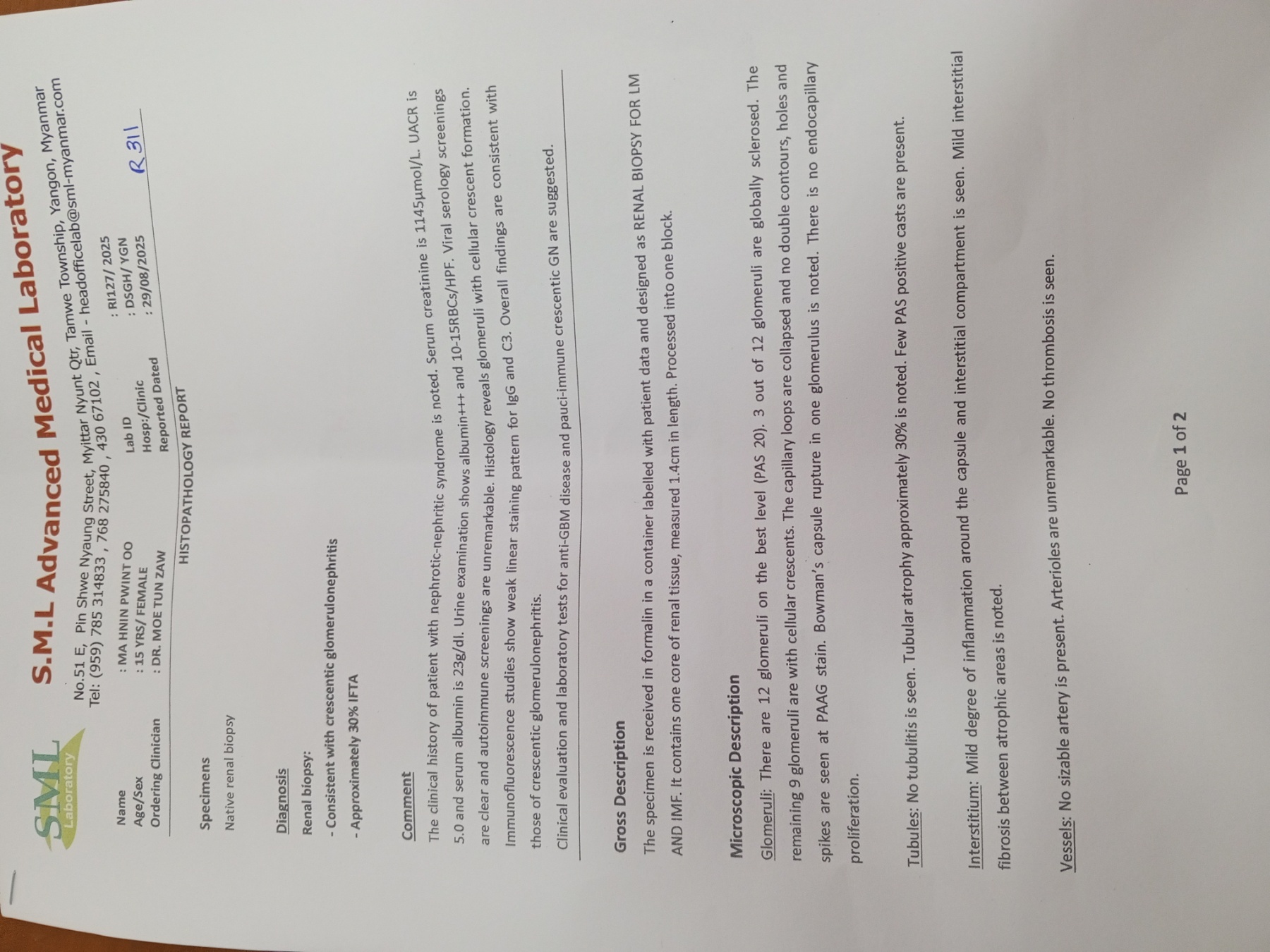
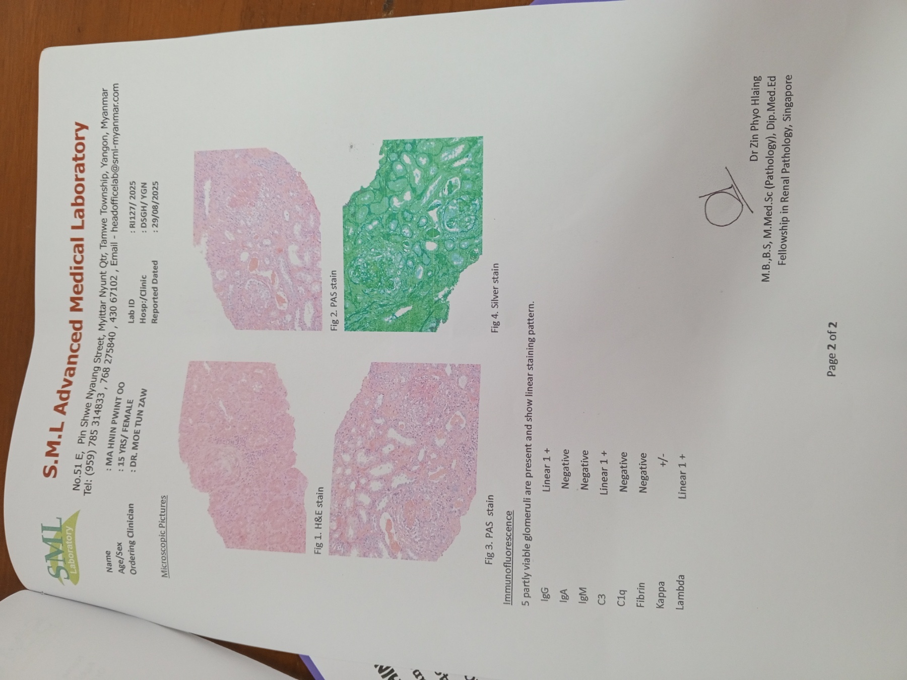
After 3 sessions of hemodialysis , her urea level-14.55 mmol/L and creatinine-554 umol/L and  renal biopsy was done with 18 G renal biopsy gun , getting 12 glomeruli , 3 out of 12 glomeruli were globally sclerosed, the remaining 9 glomeruli were with cellular crescents, IF/TA – 30 % on light microscope and Immunoflourescence microscope showing 5 partly viable glomeruli are present and show linear staining pattern (IgG Linear 1+, IgA Negative, IgM Negative, C3 Linear 1 + , C1q Negative , Fibrin Negative, Kappa +/- , Lambda Linear 1+. 

After getting renal biopsy result, IV pulse methylprednisolone 500mg+ N/S 100 ml was given for 3 days followed by oral prednisolone and IV cyclophosphamide (weight based renal friendly dosage)7.5mg/kg (IV Cyclophosphamide 300 mg + N/S 500 ml ) was given. She was planned to give 2 weekly IV cyclophosphamide for 6 dosages with the hope of dialysis independent in the future. Now she was on three times per week of maintenance hemodialysis. SEROLOGY RESULT OF ANTI-GBM ANTIBODYRENAL BIOPSY OF ANTI-GBM CASE

According to literature review, younger patients (<30 years )are more prone to pulmonary haemorrhage, and older patients (>50 years) are more prone to isolated glomerulonephritis. In contrast to literature review , this case was 15 years old girl who presented with isolated glomerulonephritis. 

Therapeutic initiation of IV immunosuppressive drugs was a little too late for this case because it took 7 days to get the serology result of anti-GBM antibody as well as renal biopsy results. Although plasmaphresis was an available treatment option to remove anti-GBM antibodies, it was too expensive to do in our resource limited country.

Kewords