TELITACICEPT TREATMENT FOR IGA NEPHROPATHY AFTER LIVER TRANSPLANTATION: A CASE REPORT


Certificate Output Instructions

For best output, select "Paper Size" as "A4" and "Margin" as "0" or "None".

To save or print to PDF, please select Print Destination > Save as PDF, enable Background Graphics under "More Settings", then click "Save".


Certificate Background


Presented the abstract " "
(Abstract co-author(s):  )


E-Poster Presentation

During the congress, E-Posters will be accessible to all participants on the congress website 24/7, as well as in the E-poster stations in the congress center. 

Preparing your E-Poster

Please review the E-Poster format requirements carefully when preparing your E-Poster. Should your E-Poster not meet the mentioned requirements, it may not be displayed as described above.

​E-Poster Submission Deadline

Please prepare and upload your E-Poster no later than March 14, 2026 11.59PM CET. After this date, you will no longer be able to prepare and upload your E-poster and it will not be displayed and accessible on the congress website.​

E-Poster Format Requirements
  • PDF file
  • Layout: Portrait (vertical orientation)
  • One page only (Dim A4: 210 x 297mm or PPT)
  • E-Poster can be prepared in PowerPoint (one (1) PowerPoint slide) but must be saved and submitted as PDF file.
  • File Size: Maximum file size is 2 Megabytes (2 MB)
  • No hyperlinks, animated images, animations, and slide transitions
  • Language: English
  • Include your abstract number
  • E-posters can include QR codes, tables and photos
https://storage.unitedwebnetwork.com/files/1288/b799da38df1c218979fa0974520f94a1.pdf
TELITACICEPT TREATMENT FOR IGA NEPHROPATHY AFTER LIVER TRANSPLANTATION: A CASE REPORT

Please follow the instructions below to input your abstract title.

Abstract titles should be brief and reflect the content of the abstract.

  • The title will not be accepted if it exceeds 25 words.
  • Type in CAPITAL LETTERS.
  • Lowercase may be used for abbreviations only, for example, mRNA.
Meixi
Wang
Jie Sheng shengjie009@126.com The Second Affiliated Hospital of Dalian Medical University Nephrology Department Dalian China -
Meixi Wang wangmeixi1114@163.com The Second Affiliated Hospital of Dalian Medical University Nephrology Department Dalian China *
-
-
-
-
-
-
-
-
-
-
-
-
-

In recent years, liver transplantation has emerged as a crucial therapeutic option for patients with end-stage liver disease. With the increasing number of individuals undergoing transplantation and the extended survival time of these patients, kidney damage following liver transplantation has gradually garnered attention. According to reports, kidney lesions after liver transplantation include various pathological types such as calcineurin inhibitors (CNIs) nephrotoxicity, IgAN, and focal segmental glomerulosclerosis (FSGS), et al. Therefore, it is crucial to promptly identify the etiology of renal impairment in patients and to implement targeted therapeutic interventions.

The pathogenesis of IgAN following liver transplantation is complex and multifactorial, involving a variety of factors including the primary liver disease prior to transplantation, hypoperfusion status during the surgical procedure, postoperative immune system reconstruction, the use of immunosuppressants, and pathogen infections. Regardless of the etiology, the pathogenesis of IgAN is considered to be inextricably linked to the immune system, with B lymphocyte activation playing an indispensable role in this process. Currently, the efficacy and safety of Telitacicept, a dual-target inhibitor of B-cell activating factor (BAFF) and a proliferation-inducing ligand (APRIL), have been confirmed in primary IgA nephropathy. However, there are no relevant reports on the application of this drug in patients following liver transplantation.

This case reports a 58-year-old male patient who was diagnosed with IgA nephropathy through kidney biopsy 9 years after liver transplantation. The patient initially received Telitacicept ( RemeGen Co., Ltd, Yantai, Shandong Province, China ) 160mg/week treatment, which was later adjusted to 240mg/week. After 7 months of treatment, the patient's proteinuria significantly improved, and kidney function remained stable. This case reveals the potential risks of IgAN after liver transplantation, emphasizing the importance of monitoring kidney function and immunological parameters in liver transplant recipients. Meanwhile, this case suggests the feasibility and effectiveness of Telitacicept in treating IgAN patients following liver transplantation, providing new insights for future therapeutic strategies.

1. Patient Admission and Clinical Background Collection

A 58-year-old Chinese male patient was admitted to our hospital due to edema and proteinuria lasting for 6 months, and a detailed collection of his clinical background was conducted. The patient had undergone an allogeneic liver transplant 9 years earlier due to chronic hepatitis B and liver cirrhosis. Postoperatively, he received long-term maintenance immunosuppressive therapy with tacrolimus, mycophenolate mofetil, and meprednisone, along with concurrent antiviral therapy for hepatitis B. Regular follow-up after the transplant showed no signs of proteinuria or renal function abnormalities. He also had a 7-year history of hypertension, with no family history of kidney disease. Six months prior to admission, the patient developed bilateral lower limb edema, and laboratory tests revealed 3+ proteinuria, prompting his admission to our department for kidney biopsy and further diagnostic examinations to clarify the underlying cause.

2. Treatment and Follow-up Protocol

After confirming the renal pathological diagnosis of IgA nephropathy (IgAN, M0E0S0T2C0), the patient was started on a targeted treatment regimen: Telitacicept at an initial dose of 160 mg per week combined with entecavir for continuous antiviral therapy. A standardized follow-up protocol was established to assess treatment efficacy and safety. Two months after the initiation of treatment, key indicators including 24-hour urinary protein excretion and serum creatinine were reassessed. Due to the lack of significant improvement in proteinuria and consideration of the patient’s relatively high baseline body mass index (BMI 29.61 kg/m²), the dose of Telitacicept was adjusted to 240 mg per week. During the follow-up period, the patient’s renal function parameters, serum immunoglobulin levels (IgA, IgM, IgG), liver function indicators (ALT, AST, LDH), and the occurrence of adverse events such as infection and hepatic injury were closely monitored to comprehensively evaluate the therapeutic effect and safety profile of the treatment.

1. Baseline Examination Findings

Physical examination of the patient revealed bilateral lower limb edema, with a blood pressure of 120/82 mmHg, heart rate of 70 beats per minute, height of 181 cm, weight of 97 kg, and a body mass index (BMI) of 29.61 kg/m². Laboratory tests (Table 1) showed the following results: renal function indicators included blood urea nitrogen 10.37 mmol/L, creatinine 186.84 μmol/L, uric acid 598.43 μmol/L, and estimated glomerular filtration rate (eGFR) 35.99 mL/min/1.73m²; 24-hour urinary protein quantification was 2.1 g/24h, and urinalysis indicated 2+ urinary protein and 0-3 red blood cells per high-power field (HP) under microscope; serum IgA was 4.02 g/L; hepatitis B virus core antibody (HBcAb) and surface antibody (HBsAb) were positive, while other hepatitis B virus markers and hepatitis B DNA were negative. Renal ultrasonography showed that both kidneys had normal size and morphology, accompanied by enhanced cortical echogenicity and indistinct corticomedullary differentiation.

Table 1 Admission hematology test data

Parameter

Value (reference range)

Hematology


White blood cell count,  109/L

5.63(3.50-9.50)

Hemoglobin, g/L

136(130-175)

Platelet count, 109/L

164(125-350)

Blood chemistry


Total protein, g/L

75.33(65-85)

Urea nitrogen, mmol/L

10.37(3.1-8.0)

Creatinine, μmol/L

186.84(57-97)

eGFR, ml/min/1.73m2

35.54(≥90)

Total cholesterol,mmol/L

4.72(2.9-5.17)

Triglyceride,mmol/L

4.08(0.22-1.7)

Parathyroid hormone,pg/ml

79.42(15-65)

Ca+,mmol/L

2.36(2.11-2.52)

IP+,mmol/L

1.08(0.85-1.51)

FK506,ng/ml

2.3

ALT, U/L

22.06(9-50)

AST, U/L

20.27(15-40)

LDH, U/L

181.87(120-250)

FBS,mmol/L

5.31(3.9-6.1)

infection


HBsAb,IU/L

53.9(0-10)

HBsAg,IU/L

0.415(0-1)

HBcAb,IU/L

0.165(>1)

HBeAb,IU/L

1.65(>1)

HBeAg,IU/L

0.109(0-1)

HBV-DNA, IU/ml

<5(0-500)

Urinalysis


protein

2+

Red blood cell, /HPF

0-1


0-2


1-3

24h UTP, mg

2093.50(0-150)


1560.01(0-150)


1625.74(0-150)

Abbreviations: eGFR, estimated glomerular filtration rate; AST, aspartate aminotransferase;ALT, alanine aminotransferase; LDH, lactate dehydrogenase; FBS, fasting blood sugar; Ig, immunoglobulin; PLA2R, phospholipase A2 receptor;MPO, myeloperoxidase; PR3, proteinase 3; ANCA, anti-neutrophil cytoplasmic antibody;UTP, urine Total Protein.

2. Renal Biopsy Pathological Results

Light microscopiy of the renal biopsy reveals(Figure 1): partial glomerular ischemic sclerosis, mild expansion of the mesangial area, and proliferation of mesangial cells with an increase in mesangial matrix. Uneven renal tubular tissue damage, observable interstitial fibrosis and tubular atrophy, focal arteriolar hyaline degeneration, thickening of interlobular arterial intima, luminal narrowing, and even occlusion. No acute pathological changes, such as crescents or collateral necrosis, were observed. Immunofluorescence showed (Figure 2) IgA(4+), IgM(3+), C3(2~3+), light chain Kappa(2+), Lambda(4+), presenting diffuse, spherical, or clustered deposits in the mesangial area. IgG, IgG1, IgG2, IgG3, IgG4, PLA2R, C1q, C4 were negative(Table 2). Immunohistochemical staining showed that HBsAg and HBcAg were negative. Electron Microscopy Examination (Figure 1) demonstrated an increase in glomerular volume. The mesangial matrix was slightly increased, with a large number of clustered electron-dense deposits in the mesangial area. Diffuse deposition of a small amount of clumped electron-dense deposits was seen in the subendothelium and subsegmental epithelium. The glomerular basement membrane showed segmental thickening (440-1300 nanometers) in areas without deposits. Local swelling of capillary endothelial cells was noted. The pathological features were consistent with IgAN (M0E0S0T2C0).

 Figure 1 Results of Renal Biopsy (A&B) Light microscope: (A) : (PASM and HE,x400) Red arrows show expansion of the mesangial area and acute tubular injury. (B) : (Masson staining,x400)   Red arrows show expansion of the mesangial area. (C&D) Electron microscopy: (C) : Red arrows denote the deposition of electron-dense material. (D) : Red arrows denote the deposition of electron-dense material.

Figure 2 Renal Biopsy Histological findings. (A-E) Histological findings.Immunofluorescence:  (A)IgA(4+),(B)IgM(3+),(C)C3(2~3+),(D)KAPPA(2+),(E)LAMDA(4+).

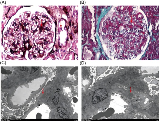
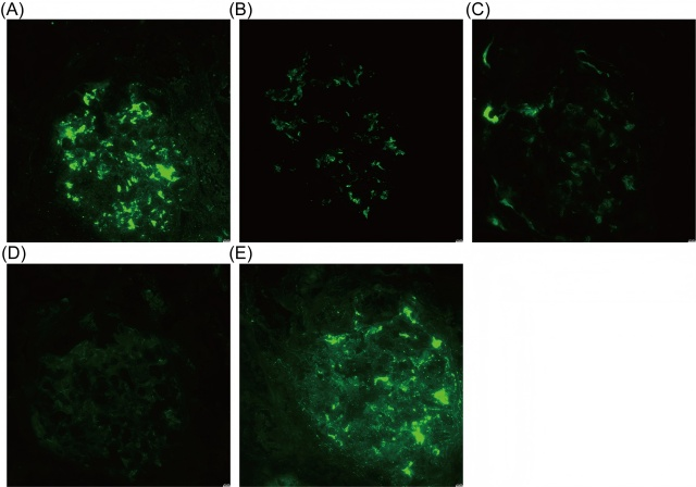
Table 2 Immunization-related examination data for admission

Parameter

Value (reference range)

IgG, g/L

12(7-16)

IgM, g/L

0.94(0.4-2.3)

IgA, g/L

4.02(0.7-4.0)

C3, g/L

1.280(0.9-1.8)

C4, g/L

0.350(0.1-0.4)

PLA2R, RU/ml

0.99(0-20)

anti-dsDNA, U/mL

5.14(0-16)

Antinuclear antibodies

normal

pANCA

normal

cANCA

normal

MPO, RU/ml

1.06(0-20)

PR3, RU/ml

3.14(0-20)

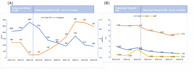
3. Treatment Outcomes

After determining the renal pathology, the patient initiated treatment with Telitacicept at a dosage of 160 mg per week and entecavir antiviral therapy. After 2 months of treatment, the 24-hour urinary protein excretion was reassessed at 2.81 g, and serum creatinine was 173μmol/L. Given the lack of significant improvement in proteinuria and considering the patient's relatively high baseline body weight (BMI 29.61 kg/m²), the dose of Telitacicept was adjusted to 240 mg per week. After 7 months of treatment, the 24-hour urinary protein excretion was reassessed at 0.90 g, serum creatinine was 203μmol/L, IgA was 1.36g/L, IgM <0.21g/L, and IgG was 8.75g/L. Meanwhile, the patient's ALT was 34.17 U/L, AST was 42.74 U/L, and LDH was 232.56 U/L, and no significant liver injury was seen. The patient achieved clinical partial remission without the occurrence of adverse events such as infection or hepatic injury. Changes in the main indicators during the patients' treatment follow-up are shown in Figures 3.


With the widespread application of liver transplantation techniques and the use of novel immunosuppressants, the survival time of patients after liver transplantation has significantly increased, leading to a corresponding rise in morbidity related to renal injury. The incidence of chronic kidney disease (CKD) following liver transplantation ranges from 17.1% to 80.0% in published studies, with a mean time to progression to end-stage renal disease (ESRD) being 66.86 months. While the traditional view holds that renal injury after liver transplantation is predominantly caused by the nephrotoxicity of CNIs, recent evidences highlight a diverse range of etiologies, including IgAN, FSGS, membranous nephropathy, membranoproliferative glomerulonephritis, and acute interstitial nephritis, et al. Therefore, early renal biopsy remains critical for etiological clarification, enabling targeted immunosuppressive regimens and primary disease management.

The present case exhibited biopsy-confirmed IgAN. A retrospective study in China identified IgAN in 2 of 14 patients (14.3%) undergoing post-transplant renal biopsy, while Choudhary et al. reported IgAN in 4 of 26 patients (15.4%) with refractory post-transplant renal insufficiency. Secondary glomerular IgA deposition is linked to multiple systemic disorders, most frequently hepatic IgAN. Hepatic IgAN typically follows an indolent course due to impaired hepatic IgA clearance and portal shunting mechanisms. Although post-transplant resolution is uncommon, prognosis remains favorable. In this patient, prolonged hepatitis B stability and preserved liver function suggested primary IgAN rather than secondary forms.

The pathogenesis of IgAN involves the synthesis of galactose-deficient IgA1 (Gd-IgA1), which forms immune complexes that deposit in renal mesangium, triggering mesangial cell proliferation, extracellular matrix expansion, cytokine/chemokine release, and activation of the complement pathway. B-cell dysregulation drives Gd-IgA1 and anti-Gd-IgA1 autoantibody production, mediated by overexpression of BAFF and APRIL. These cytokines critically regulate B-cell maturation, class-switching, and antigen-specific plasma cell survival.  However, detection of Gd-IgA1 in IgAN is complex and unstable, so we did not use Gd-IgA1 as a basis for evaluating this patient's condition.

Telitacicept, a dual BAFF/APRIL inhibitor, suppresses B-cell maturation and plasma cell viability, halving circulating Gd-IgA1 and IgG immune complex levels in IgAN patients. Prior studies corroborate its safety in transplant settings, showing chronic rejection mitigation and allograft survival extension in cardiac transplantation. A phase II trial demonstrated superior efficacy with telitacicept 240 mg/week compared to 160 mg/week, with comparable adverse event rates. Given the patient’s elevated BMI (29.61 kg/m²), we escalated dosing to 240 mg/week, achieving partial clinical remission (24-hour urinary protein: 0.90 g) after 7 months. 

In this case, proteinuria decreased significantly after treatment with telitacicept 240 mg/week, renal function, IgG level and hepatic function were stable, and no adverse events such as hepatitis B reactivation and infection occurred during the follow-up period. This suggests that targeted therapies represents a promising strategy for IgAN after liver transplantation, warranting further investigation through multicenter trials.

Kewords