TUBERCULOSIS-INDUCED HYPERCALCEMIA IN PREVIOUS STAGE 5 CKD HYPOCALCEMIA: A COMPLEX DIAGNOSTIC DILEMMA

https://storage.unitedwebnetwork.com/files/1099/948b9d25e78ccc2d63b8dc81baf65889.pdf
TUBERCULOSIS-INDUCED HYPERCALCEMIA IN PREVIOUS STAGE 5 CKD HYPOCALCEMIA: A COMPLEX DIAGNOSTIC DILEMMA
Faviola Marie
Negrón Alick
Andres Aranda - G. de Quevedo dr.andresaranda@gmail.com Universidad Autónoma de Guadalajara / Hospital General de Occidente School of Medicine / Nephrology Department Zapopan
Linda Giovanna Ruiz - Fabian dra.giovannaruiz@gmail.com Universidad Autónoma de Guadalajara School of Medicine Zapopan
Hector Enrique Garcia - Bejarano garciabejarano.garcia@gmail.com Hospital General de Occidente Nephrology Department Zapopan
Erick Ivan Marrero - Santiago erick.marrero@edu.uag.mx Universidad Autónoma de Guadalajara School of Medicine School of Medicine Zapopan
David Pagan - Maldonado david.pagan@edu.uag.mx Universidad Autónoma de Guadalajara School of Medicine School of Medicine Zapopan
Cristian Xavier Rivera - Benitez cristian.rivera@edu.uag.mx Universidad Autónoma de Guadalajara School of Medi School of Medicine Zapopan
Valeria Mora - Castillo vmora@edu.uag.mx Universidad Autónoma de Guadalajara School of Medi School of Medicine Zapopan
Kashif Adil Ahmad kashif@edu.uag.mx Universidad Autónoma de Guadalajara School of Medi School of Medicine Zapopan
Miranda Robledo - Amezcua miranda.robledo@edu.uag.mx Universidad Autónoma de Guadalajara School of Medi School of Medicine Zapopan
Alejandra Mariel Franco - Martinez alejandram@edu.uag.mx Universidad Autónoma de Guadalajara School of Medi School of Medicine Zapopan
Enid Duran - Gonzalez enid.duran@edu.uag.mx Universidad Autónoma de Guadalajara School of Medi School of Medicine Zapopan
Simon Quetzalcoalt Rodriguez - Lara simon.rodriguez@edu.uag.mx Universidad Autónoma de Guadalajara School of Medi School of Medicine Zapopan
Javier Adan Castañeda - Moreno javier.adan.castaneda@edu.uag.mx Universidad Autónoma de Guadalajara School of Medi School of Medicine Zapopan
Lucia Elizabeth Alvarez - Palazuelos lucia.palazuelos@edu.uag.mx Universidad Autónoma de Guadalajara School of Medi School of Medicine Zapopan
Miguel Alejandro Davalos - Benitez miguel.davalos@edu.uag.mx Universidad Autónoma de Guadalajara School of Medi School of Medicine Zapopan

Parathyroid hormone (PTH) and vitamin D play important roles in maintaining calcium balance. PTH contributes to the increase of serum calcium levels by promoting the release of calcium from bones, increasing calcium reabsorption in the kidneys, and stimulating the activity of 1-alpha-hydroxylase, which converts inactive vitamin D (25-hydroxy vitamin D) into its active form, calcitriol (1,25-dihydroxy vitamin D). Reduced synthesis of the enzyme 1-alpha-hydroxylase is noted in Chronic Kidney Disease, decreasing the presence of calcium in serum, and by entering in a hyper parathyroid state, the body tries to compensate for the hypocalcemia. Patients with CKD are more susceptible to TB infection or reactivation of latent TB infection (LTBI) due to their immunosuppression. Granulomatous diseases, such as tuberculosis, can disrupt the calcium-phosphate homeostasis leading to hypercalcemia independently of PTH levels by promoting the overproduction of calcitriol within granulomas due to activated macrophages that express 1α-hydroxylase. In some reports, the incidence of hypercalcemia in patients with TB has been reported to from 2.3 up 28 percent. Calcitriol plays a crucial role in the regulation of granulomatous inflammation and influences the cell-mediated immunity in tuberculosis. Most TB patients present hypocalcemia, and if presented with hypercalcemia, this manifestation is usually asymptomatic.


We present a case of a 21-year-old female diagnosed with end-stage chronic kidney disease (CKD) of unknown etiology in Guadalajara, Jalisco, Mexico, a city known for its high incidence of CKD at an early age. She had no relevant, known personal medical history or chronic conditions that could have been attributed to the precise etiology of chronic kidney disease. 


However, she had a chronic habit of using crystal meth, averaging around 5 grams per day over the last year, which could have exacerbated an underlying cause. Additionally, she was a heavy tobacco user, starting at the age of 15, smoking up to 60 cigarettes a day, with a tobacco index of 18, putting her at moderate risk for chronic obstructive lung disease.


The patient was not living with her family and had a limited social and family support network. She was first admitted in November 2022 after experiencing a chronic episode of diarrhea and abdominal pain. Initially, she sought ambulatory care from general practitioners, who prescribed symptomatic treatment. However, after nearly one and a half months, her pain progressed to become unbearable, accompanied by nausea, vomiting, and fever. She sought medical attention again, and laboratory tests revealed the presence of azotemia, leading to her referral to a secondary-level hospital. 


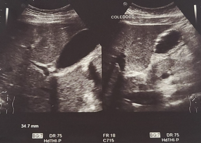
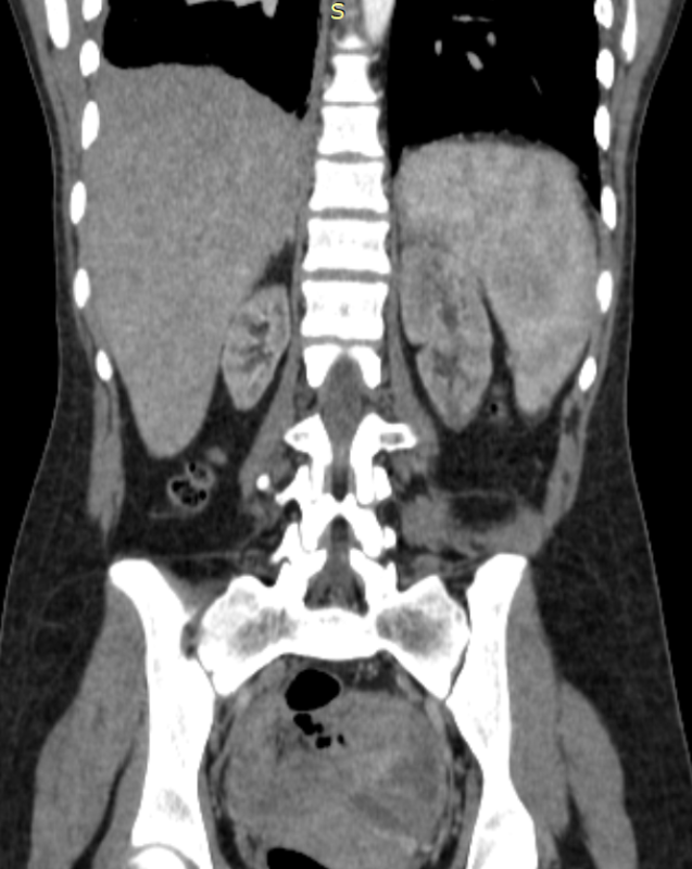
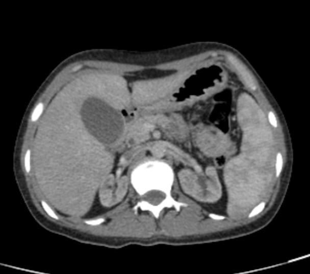
Upon admission, she was evaluated for acute abdominal pain attributed to an acute episode of pancreatitis. An abdominal CT scan was performed, revealing hepatomegaly, bilateral renal hypoplasia, and changes in the cortex/medulla ratio. An abdominal ultrasound showed the presence of an acoustic shadow, suggesting the presence of choledocholithiasis (image 1). Subsequently, a cholangiography resonance scan found that the stones were not obstructing the biliary tract. She received symptomatic treatment to manage gastrointestinal symptoms and was diagnosed with acute kidney injury superimposed on her chronic kidney disease due to an acute episode of biliary pancreatitis. 


Despite several days of in-hospital treatment, her azotemia persisted. Laboratory results showed metabolic abnormalities, such as hypocalcemia and azotemia, in conjunction with the imaging findings. She was ultimately diagnosed with end-stage chronic kidney disease after assessment by the nephrology team. She was prescribed predialysis conservative treatment, including bicarbonate, blood pressure control with ACE inhibitors, and diuretics.

The patient was assessed by the nephrology team, and renal replacement therapy was offered to her with hemodialysis. However, the patient declined this treatment and requested voluntary discharge, despite being informed of the risks associated with this decision. Pre-dialysis treatment measures were provided to her to continue her outpatient management and evaluation.


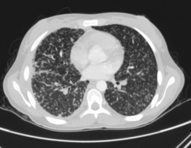
Unfortunately, the patient did not follow the discharge instructions and abandoned her treatment. Two months later, in January 2023, the patient returned to the hospital due to an episode of progressive productive cough with yellowish sputum that persisted throughout the day, accompanied by dyspnea that progressed from exertional to resting dyspnea and orthopnea. She also experienced high-grade intermittent fever and night sweats. X-ray and thoracic and abdominal CT scan images (image 2) suggested an active Mycobacterium tuberculosis lung infection, but not imaging suggesting urinary tract involvement. There was no presence of gallstones in the gallbladder. Even though the three serial Acid-Fast Bacilli (AFB) tests came back negative, she was diagnosed with TB by the infectious disease department. 


It came to our attention that, despite not following any of the nephrology team's recommendations and discontinuing all prescribed treatment, she had not received any calcium or other related treatments for mineral bone disorders. She was admitted without symptoms of severely elevated levels of ionized calcium (12.5 mg/dL). Levels of PTH (7.50) and Vitamin D (9.0) (TABLE 1) were requested just before we requested the level of calcitriol; however, the patient escaped from the hospital.


In April 2023, the patient returned to the ED due to persistent signs of active pneumonia, such as dyspnea and a productive cough. She also exhibited symptoms of uremia, including pruritus, nausea, and hyporexia, along with two days of evolving epigastric pain radiating to the left upper quadrants. Her lipase levels were elevated to three times the upper high limit, and her serum calcium levels measured 10.9 with an ionized calcium level of 1.39. She was diagnosed with a new episode of acute pancreatitis. A new POCUS abdominal ultrasound revealed no abnormal findings in the hepatic and biliary tracts. She received analgesics, IV fluids, and pre-dialytic treatment, and her abdominal symptoms improved.


Once again, she was offered renal replacement therapy and TB treatment, but she declined and requested voluntary discharge. As of the time of writing this text, she has not returned to the hospital.

Hypocalcemia is one of the electrolyte imbalances in patients with advanced CKD, as the patient initially presented. It's worth noting that, despite having an active pulmonary TB, the patient experienced two episodes of severe hypercalcemia and suppressed PTH levels. Unfortunately, we didn't have the opportunity to obtain serum Calcitriol levels. During her last hospitalization, the patient met 2 out of 3 criteria for acute pancreatitis, with no suggestive evidence of biliary involvement. There's a possibility that hypercalcemia might have triggered this second event. This case underscores the importance of addressing calcium disorders thoroughly and highlights that even in cases of end-stage CKD, PTH can respond normally and be suppressed in instances of hypercalcemia.

E-Poster Format Requirements
  • PDF file
  • Layout: Portrait (vertical orientation)
  • One page only (Dim A4: 210 x 297mm or PPT)
  • E-Poster can be prepared in PowerPoint (one (1) PowerPoint slide) but must be saved and submitted as PDF file.
  • File Size: Maximum file size is 2 Megabytes (2 MB)
  • No hyperlinks, animated images, animations, and slide transitions
  • Language: English
  • Include your abstract number
  • E-posters can include QR codes, tables and photos